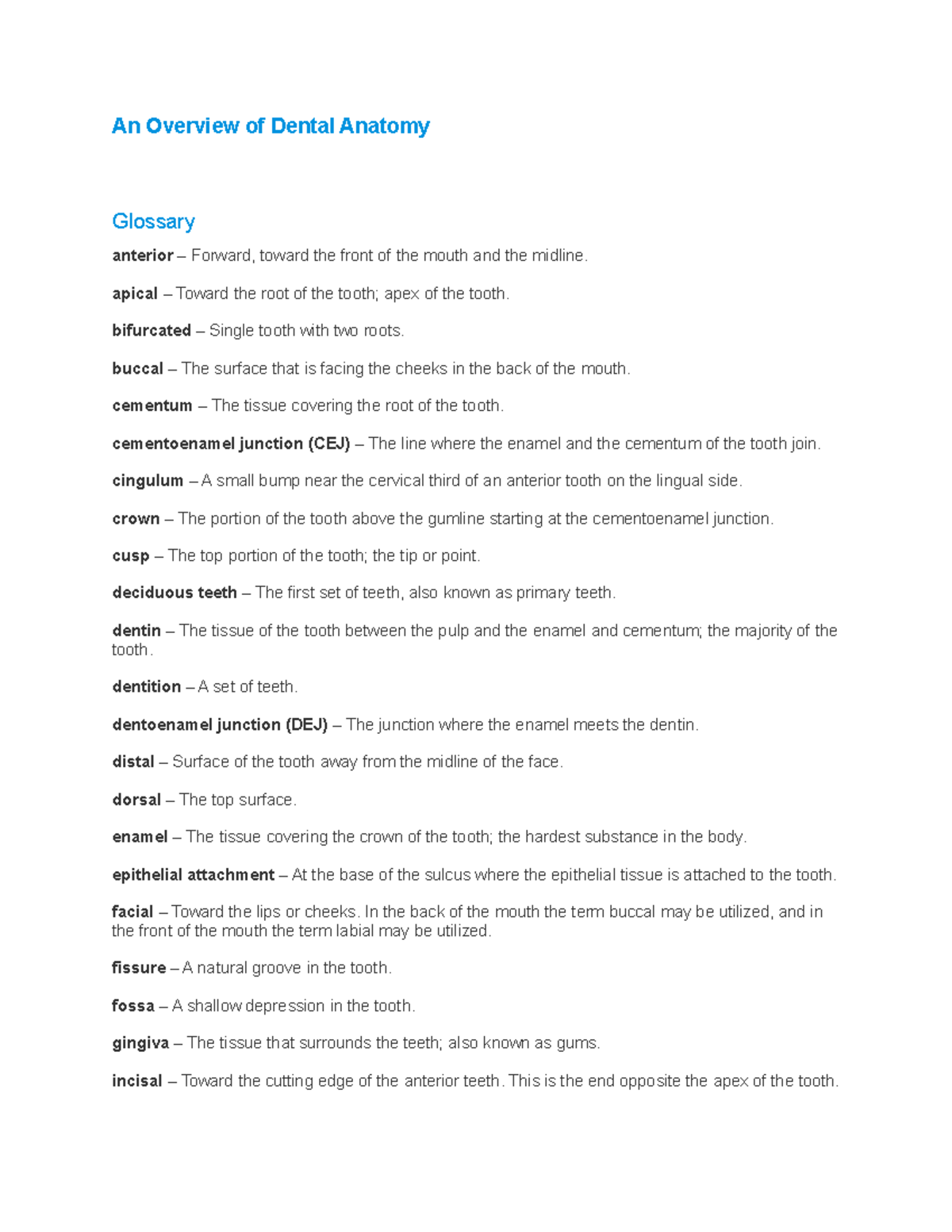
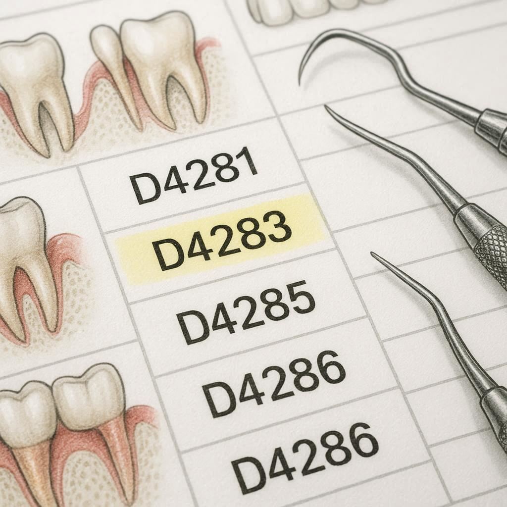
Understanding Dental Anatomy and Terminology: A Complete Guide
When you sit in the dentist’s chair, the words you hear—incisor, pulp chamber, occlusal—can feel like a foreign language. Decoding dental anatomy and terminology not only demystifies the experience but also empowers you to make better oral‑health decisions. Below, we break down the structures of a tooth, translate the most common jargon, and explain why that knowledge matters for everyday care.
Key Structures of the Tooth
A tooth isn’t just a single piece of enamel; it’s a complex assembly of hard and soft tissues, each with a specific role. Understanding these parts helps you visualize what’s happening during a filling, root canal, or routine cleaning.
Enamel
The outermost layer, enamel, is the hardest substance in the human body. It shields the inner dentin from the wear and tear of chewing. Because enamel can’t regenerate, protecting it with fluoride and proper brushing is crucial.
Dentin
Just beneath the enamel lies dentin, a porous, yellowish tissue that makes up the bulk of the tooth. Dentin transmits sensations to the pulp, so when it becomes exposed—say, from a cavity—you may feel heightened sensitivity.
Pulp Chamber and Root Canal
The pulp chamber houses nerves, blood vessels, and connective tissue. From this chamber extend the root canals, which travel down the roots to supply the tooth with nutrients. When infection reaches the pulp, a root‑canal treatment removes the compromised tissue and seals the space.
Cementum
Covering the root surface is cementum, a bone‑like material that anchors the tooth to the periodontal ligament. Though thinner than enamel, cementum plays a vital role in tooth stability.
Periodontal Ligament (PDL)
The PDL is a network of collagen fibers that connect cementum to the surrounding alveolar bone. It acts like a shock absorber, allowing slight tooth movement under pressure and supporting overall bite dynamics.
Alveolar Bone
This is the jawbone that houses the tooth sockets. Healthy alveolar bone is essential for long‑term tooth retention; bone loss can lead to loosening or even tooth loss.
Common Terminology Explained
Dental professionals use a concise lexicon to describe conditions, procedures, and anatomy. Below is a quick reference you’ll likely encounter on a dental chart or during a consultation.
- Incisor – The front teeth designed for cutting. Humans have eight incisors, four on the top and four on the bottom.
- Canine – Also called cuspids; these pointed teeth tear food and guide the bite.
- Premolar – Located behind canines, premolars have broader surfaces for crushing.
- Molars – The largest teeth, situated at the back, responsible for grinding.
- Occlusal – Refers to the chewing surface of posterior teeth.
- Proximal – The side surfaces of a tooth that face neighboring teeth; these areas are common sites for caries.
- Restoration – Any procedure that restores a tooth’s form or function, such as fillings, crowns, or veneers.
- Extraction – The removal of a tooth, either simple (visible) or surgical (impacted).
- Periodontitis – Advanced gum disease that destroys the supporting bone and connective tissue.
- Prophylaxis – Professional cleaning aimed at preventing disease, often called a “prophy.”
Familiarity with these terms reduces anxiety; you’ll know whether a dentist is suggesting a routine cleaning or a more involved procedure.
Why Knowing Dental Anatomy Matters
Beyond the jargon, understanding the anatomy can shape how you protect your smile.
First, it clarifies why certain habits are harmful. For instance, chewing ice can fracture enamel, while aggressive brushing can wear away cementum at the gum line, exposing dentin and causing recession.
Second, it informs treatment choices. When a dentist recommends a crown, you’ll recognize that the underlying tooth structure—perhaps heavily damaged dentin—needs that extra reinforcement.
Finally, it aids communication with your oral‑health team. Asking, “Is the decay reaching the pulp chamber?” shows you’re engaged and helps the dentist explain the urgency of a root canal versus a simple filling.
Frequently Asked Questions
What is the difference between a crown and a veneer?
A crown encircles the entire tooth, restoring both function and appearance, typically used when substantial structure is lost. A veneer is a thin shell placed on the front surface, primarily for cosmetic improvement, and requires less tooth reduction.
How can I protect my enamel?
Limit acidic foods and drinks, use a fluoride toothpaste, and avoid brushing immediately after consuming citrus or soda—wait at least 30 minutes to give saliva a chance to neutralize acids.
Is a root canal painful?
The procedure itself is performed under local anesthesia, so you shouldn’t feel pain during it. Post‑treatment soreness is common but usually manageable with over‑the‑counter pain relievers.
Can gum disease affect the bone?
Yes. In periodontitis, bacterial toxins and inflammation gradually destroy the alveolar bone, leading to tooth mobility and potential loss if untreated.